To avoid a lead apron artifact on a panoramic radiograph is a crucial aspect of dental imaging, ensuring accurate and reliable diagnostic information. This comprehensive guide delves into the formation, appearance, and clinical significance of lead apron artifacts, providing practical methods to minimize their occurrence and optimize image quality.
Proper positioning of the lead apron, utilization of additional shielding materials, and optimization of exposure parameters play a vital role in reducing scatter radiation and preventing artifacts. Patient education and clear communication are equally important, emphasizing the significance of cooperation and understanding the instructions provided before the radiographic examination.
Lead Apron Artifact on Panoramic Radiographs: To Avoid A Lead Apron Artifact On A Panoramic Radiograph

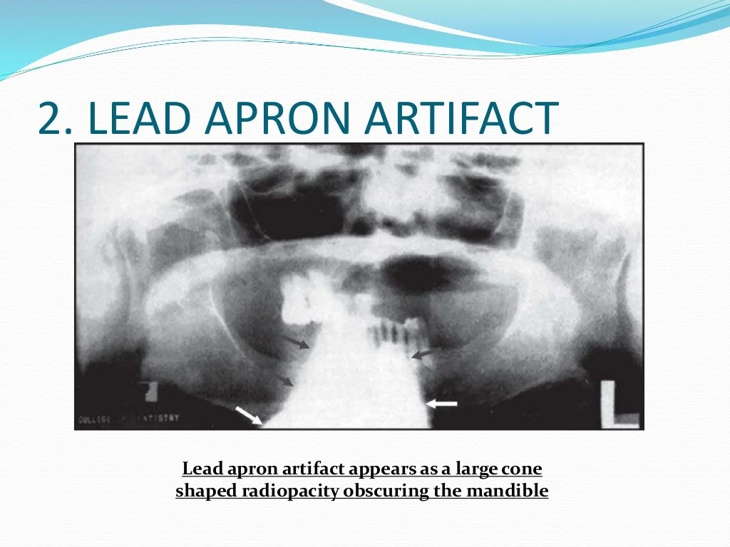
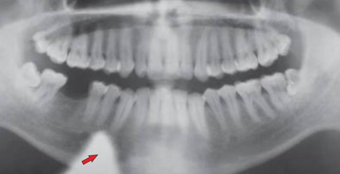
Lead apron artifacts are a common problem in panoramic radiography, affecting the diagnostic quality of the images. They are caused by the scattering of X-rays by the lead apron worn by the patient to protect their body from radiation exposure.
The characteristic appearance of lead apron artifacts is a dark band or streak across the panoramic image, typically in the region of the thyroid gland or mandibular symphysis. The artifact can obscure underlying anatomical structures, making it difficult to interpret the radiograph.
Lead apron artifacts can have clinical significance, as they can lead to misdiagnosis or missed diagnoses. For example, a dark band across the thyroid gland may obscure a thyroid nodule or mass, potentially leading to a delay in diagnosis and treatment.
Methods to Avoid Lead Apron Artifacts
There are several methods that can be employed to minimize or eliminate lead apron artifacts on panoramic radiographs.
Proper Positioning of the Lead Apron
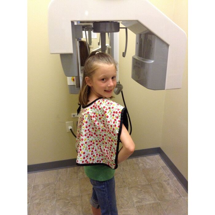
The lead apron should be positioned correctly to ensure that the thyroid gland and mandibular symphysis are not exposed to scattered radiation. The apron should be placed over the shoulders and extend down to the waist, with the edges of the apron overlapping at the back.
Use of Additional Shielding Materials
Additional shielding materials, such as thyroid collars or bismuth shields, can be used to further reduce scatter radiation and minimize lead apron artifacts. Thyroid collars are placed around the patient’s neck to protect the thyroid gland, while bismuth shields can be placed over the mandibular symphysis.
Optimization of Exposure Parameters
Optimizing exposure parameters, such as kVp and mAs, can help to reduce lead apron artifacts. Lower kVp settings and shorter exposure times can minimize scatter radiation and improve image quality.
Patient Education and Communication, To avoid a lead apron artifact on a panoramic radiograph
Patient cooperation is essential in avoiding lead apron artifacts. Patients should be instructed to remain still and avoid moving during the radiographic exposure. They should also be informed about the importance of wearing the lead apron correctly.
Visual aids or demonstrations can be used to enhance patient understanding and ensure proper lead apron positioning. A simple diagram or demonstration can help patients visualize the correct position of the apron and the potential consequences of improper positioning.
Alternative Imaging Techniques
In some cases, alternative imaging techniques may be used to minimize or eliminate lead apron artifacts. These techniques include:
- Cone beam computed tomography (CBCT): CBCT uses a cone-shaped X-ray beam to create three-dimensional images of the maxillofacial region. This technique reduces scatter radiation and eliminates the need for a lead apron.
- Digital panoramic radiography: Digital panoramic radiography uses a digital sensor to capture the panoramic image. This technique also reduces scatter radiation and minimizes the risk of lead apron artifacts.
The choice of alternative imaging technique depends on the clinical situation and the specific needs of the patient.
Quality Assurance and Monitoring
Quality assurance measures are essential to minimize lead apron artifacts and ensure the production of high-quality panoramic radiographs. These measures include:
- Image review: Radiographs should be reviewed for the presence of lead apron artifacts. Any artifacts should be identified and corrected through appropriate measures.
- Artifact assessment: The severity of lead apron artifacts can be assessed using objective criteria, such as the artifact index. This index measures the darkness and width of the artifact and can be used to evaluate the effectiveness of artifact reduction techniques.
- Phantom studies: Phantom studies can be used to evaluate the effectiveness of different artifact reduction techniques. Phantoms simulate the human body and can be used to assess the amount of scatter radiation and the resulting artifact.
Regular quality assurance measures help to ensure that lead apron artifacts are minimized and that panoramic radiographs are of high diagnostic quality.
FAQ Explained
What is a lead apron artifact?
A lead apron artifact is a radiographic artifact caused by the lead apron worn by the patient during panoramic radiography, resulting in a dark band or shadow on the image.
How can lead apron artifacts be avoided?
Lead apron artifacts can be avoided by proper positioning of the apron, ensuring it is placed below the chin and not overlapping the area of interest, and by using additional shielding materials, such as thyroid collars, to minimize scatter radiation.
Why is it important to avoid lead apron artifacts?
Lead apron artifacts can obscure anatomical structures, potentially leading to misinterpretation and inaccurate diagnoses.

